Sheep Eye Anatomy
Sheep eye anatomy. Virtual sheep eye dissection. A Gross Histologic and Histochemical Study of the Eye Adnexa of Sheep and Goats-Ram Das Sinha 1965 Fetal Pig Dissection Guide- 1993 Clinical Anatomy and Physiology Laboratory Manual for Veterinary Technicians-Thomas P. Log In Sign Up.
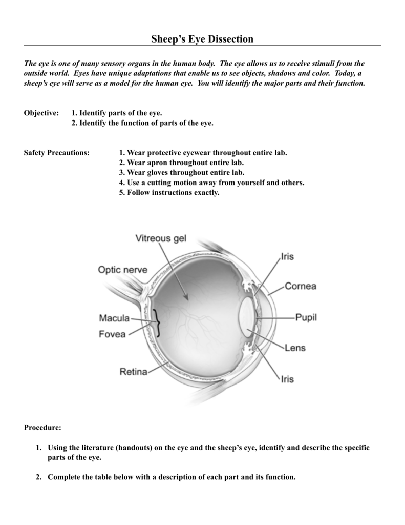
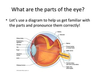
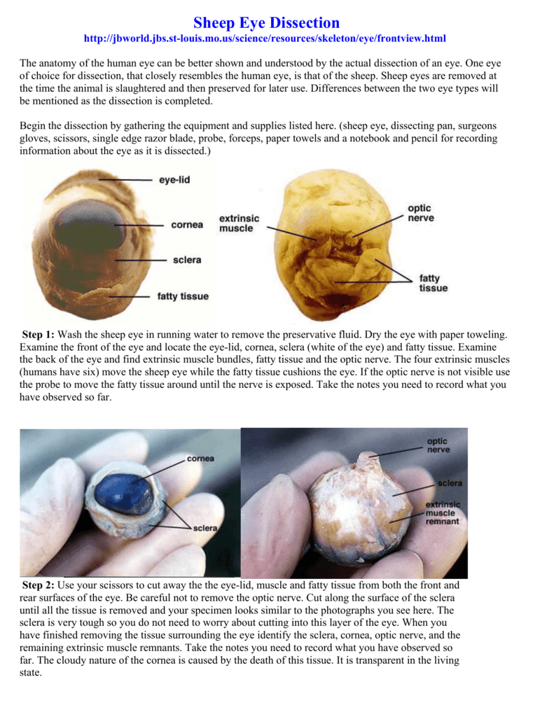
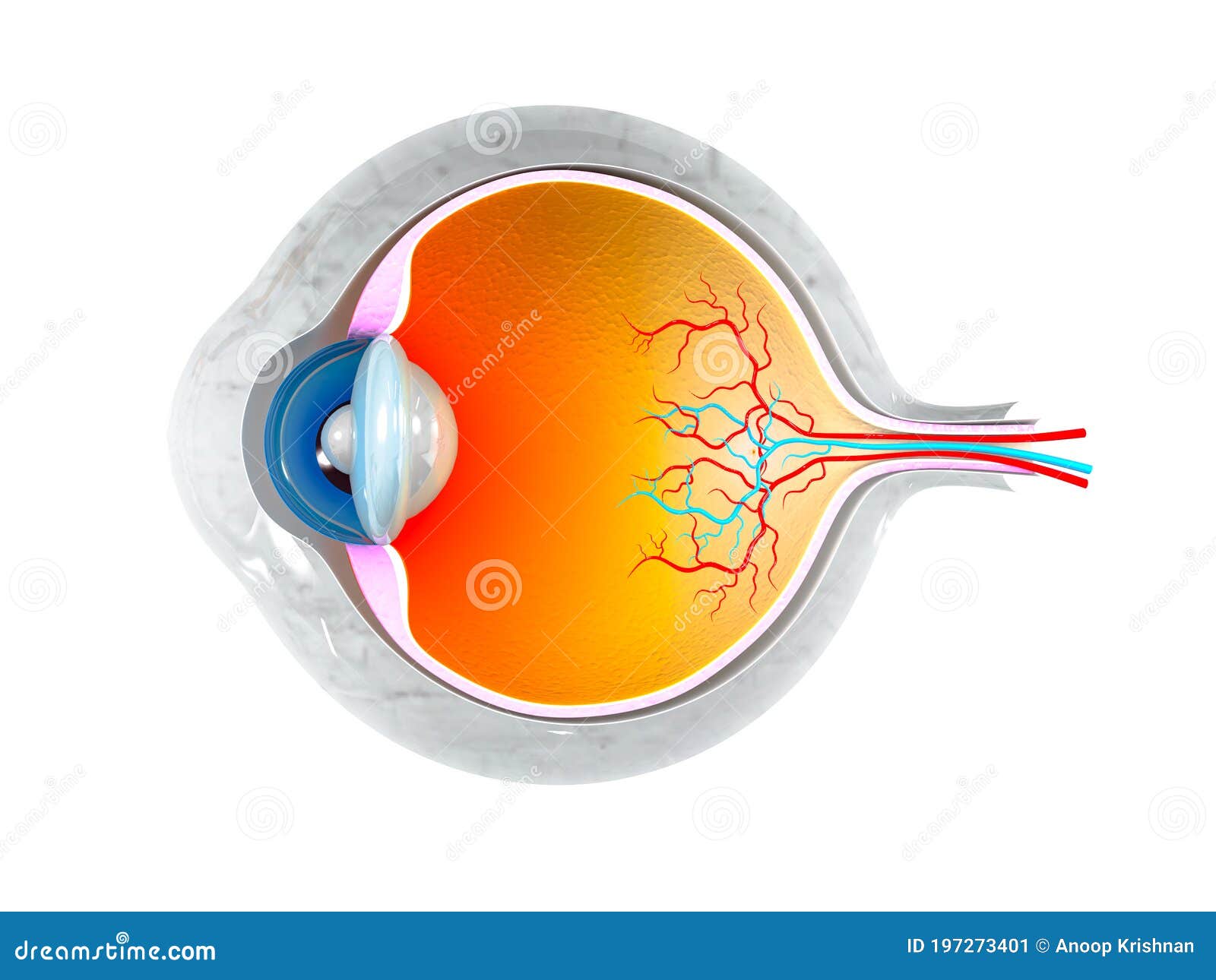
Label the eye diagram. Wash the sheep eye in running water to remove the preservative fluid. Test your knowledge of the sheep eye.
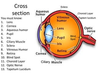
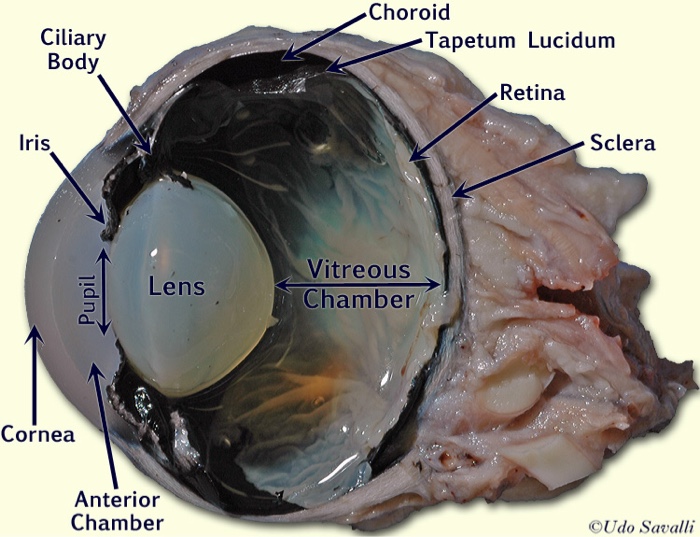
Sheep eye has tapectum lucidum that is a layer f tissues causing reflection of light. The clear dome-shaped surface that covers the front of the eye. Posted by 6 minutes ago.
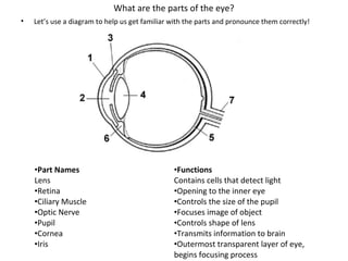
The opening through which light enters the eye. It is located in the center of the retina. The lens is a clear part of the eye behind the iris that helps to focus light or an image on the retina.
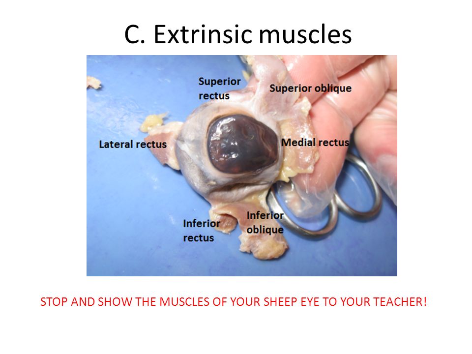
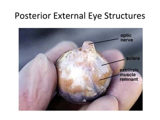
Print a Diagram of the Eye - Click on this link and then use the browser print command to produce a diagram to use with the student tutorial. Differences between the two eye types will be mentioned as the dissection is completed. Examine the back of the eye and find extrinsic muscle bundles fatty tissue and the optic nerve.
Sheep eye is placed sideways on its head whereas humans have forward facing eyes. Tough white outer coat of the eyeball. These muscles move the eyeball in its socket from.
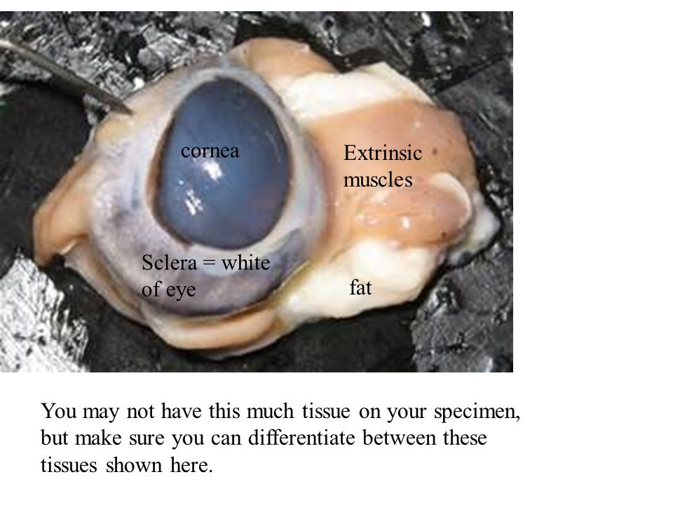
The sclera is the tough outer coat of the eyeball which helps keep the shape of the eyeand protect it from injuryThe pinkish parts around the sclera are the remnants of extrinsic muscles which have been cut off. Human eye has six muscles for eye.
Examine the back of the eye and find extrinsic muscle bundles fatty tissue and the optic nerve.
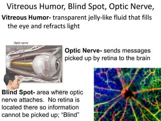
Recommend all students watch this video prior to attending eye dissecti. Examine the back of the eye and find extrinsic muscle bundles fatty tissue and the optic nerve. A tough clear covering over the iris and the pupil that helps protect the eye. The colored part of the eye. Press J to jump to the feed. Examine the front of the eye and locate the eye-lid cornea sclera white of the eye and fatty tissue. The macula is the small sensitive area of the retina that gives central vision. To study the structure of the mammalian eye and relate its structure to the process of vision. While human eye has circular pupil sheep eye has oval shaped pupil.
A Clinical Laboratory Manual 2E is designed as a lab manual for your veterinary technology and pre-veterinary medicine students who possess a basic knowledge of biology. Human eye has six muscles for eye. The colored part of the eye. Press J to jump to the feed. To study the structure of the mammalian eye and relate its structure to the process of vision. Test your knowledge of the sheep eye. The goat and sheeps eye is similar to a human eye with a lens cornea iris.


















Post a Comment for "Sheep Eye Anatomy"